

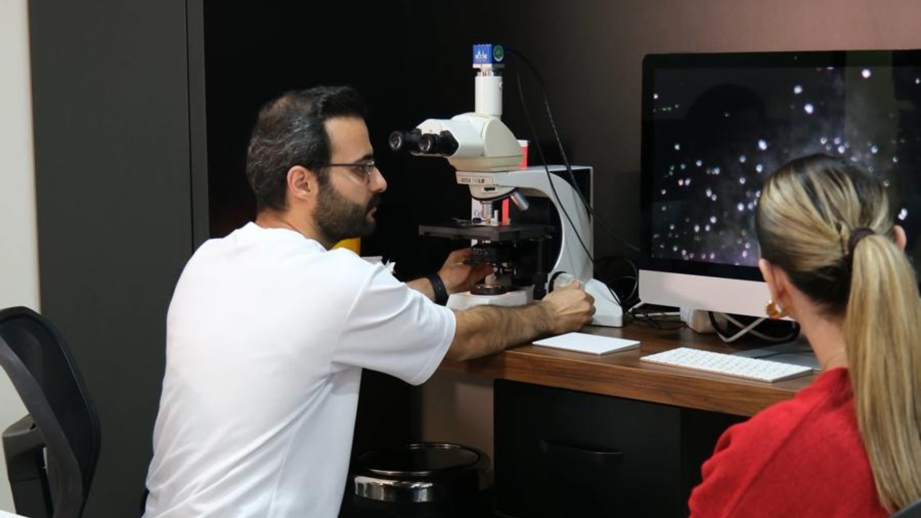
Principle of Darkfield Microscopy
Darkfield microscopy is a specialised technique that allows for detailed examination of biological samples. It illuminates the sample from the sides, creating a dark background, which makes the cells and microorganisms appear brighter and clearer. This method is particularly suited for observing thin and transparent structures.
The core principle of darkfield microscopy involves collecting light that is reflected or scattered by the sample. Unlike standard microscopes, where light is directed straight onto the sample, darkfield microscopy uses light coming from around the sample, capturing only the scattered light with the objective lens. This results in a bright sample against a dark background.
This technique is frequently used in live blood analysis, enabling the observation of cells and microorganisms in their natural state. Such analyses provide valuable insights for research and educational purposes, especially for guests. For instance, in scientific studies, darkfield microscopy is a powerful tool for examining cell dynamics and microorganism behaviours.
While darkfield microscopy offers significant visual data for informative processes, it does have limitations. The thickness and opacity of samples can impact image quality, and sometimes, additional microscopy methods are required for a more comprehensive analysis.
What Do Participants See?
Darkfield microscopy enables detailed observation of cells and microorganisms, making it ideal for clearly viewing cell shapes and movements. Participants can witness live images of blood cells and other microscopic structures, revealing interactions, structures, and movement capabilities.
For guests, this experience provides a unique opportunity to delve into the microscopic world. Darkfield microscopy allows observation of cellular behaviour in natural environments, offering valuable insights for those interested in biology and medicine. Understanding the healthy or pathological states of cells becomes more accessible with this technique.
The visuals obtained through darkfield microscopy enhance comprehension of the microscopic world. For example, cell interactions or microorganism movements become more understandable. Within this context, the 7-day doctor-supervised program at Cellavia × Kilikya Palace in Göynük offers participants the chance to make such observations.
This technique significantly aids clients in exploring and understanding the complex microscopic world. Participants can deepen their scientific observations and better grasp the dynamics at play.
Click here to learn more about science.Diagnostic Capabilities of the Device
Darkfield microscopy allows for detailed examination of cell structures and movements. However, its diagnostic capabilities are limited and not suitable for direct medical diagnosis. While ideal for observing blood cell morphology and movements, these observations do not provide sufficient data for clinical decisions.
The microscope is primarily used to study the structural features and movements of blood cells, offering insights into their health and general conditions. However, this information is observational and not suitable for diagnosing health conditions. For instance, while it provides data on blood cell movement and shape, it cannot confirm the presence or absence of a disease.
Darkfield microscopy is more commonly employed for educational purposes and research environments. Guests can gain in-depth knowledge about cell biology and microorganism behaviour. However, its use in clinical applications and diagnostic processes is not advisable.
In summary, darkfield microscopy is an effective tool for observing cell structure and behaviour but should not be used for medical diagnosis. For more information, visit our science page.
Informative Boundaries and Safety
Darkfield microscopy holds great potential in educational and research settings. However, certain boundaries and safety measures must be adhered to for safe and effective use. This complex optical system should only be operated by trained individuals to ensure user safety and data accuracy.
In terms of informative boundaries, darkfield microscopy is generally used in biology and medical knowledge areas. Users must understand the device's working principles, optical settings, and data interpretation to prevent misuse and fully leverage its advantages.
From a safety standpoint, precautions are necessary to protect the microscope's optical components and users' eye health. The use of personal protective equipment is recommended when operating the microscope.
Designed for educational and research use, darkfield microscopy is not recommended for commercial or clinical applications. Such devices should be used under trained supervision and within specific protocols. For instance, doctor-supervised programs can ensure correct and safe microscope use. For more information, visit our science page.
Use at Cellavia Measurement Table
Darkfield microscopy is frequently employed in educational programs, allowing guests to observe biological structures and cellular phenomena in detail. It plays a crucial role in scientific research methods and measurement sessions.
The Cellavia Measurement Table facilitates the effective use of darkfield microscopy, helping guests develop measurement discipline and enhance their observation skills. The detailed images provided by the microscope make understanding cellular dynamics and interactions easier, fostering a deeper comprehension of biological processes.
Doctor-supervised programs, such as the 7-day sessions at Kilikya Palace in Göynük, enhance the educational potential of darkfield microscopy. These programs offer practical experience in microscope use, combining theory with practice and developing scientific research skills.
The data obtained through darkfield microscopy aids in understanding biological structures, making educational programs more effective and enhancing guests' scientific thinking skills. The Cellavia Measurement Table is a vital tool that supports this process and plays a key role in achieving educational goals.
Data Obtained with Darkfield Microscopy
Darkfield microscopy is a valuable tool for examining biological samples, providing detailed information about cell morphology. It allows for clearer observation of structural differences and organisation, offering valuable insights for researchers in cell biology and pathology.
Additionally, darkfield microscopy is used to observe microorganism movements, particularly suitable for examining live bacteria and protozoa. Understanding microorganism movement patterns and behaviours contributes to biological process comprehension, crucial for microbiology research and educational material development.
Darkfield microscopy serves as an educational resource, helping guests better understand cell and microorganism structures. In educational programs, especially those under doctor supervision, darkfield microscopy applications provide practical experience. For instance, doctor-supervised 7-day programs at Cellavia × Kilikya Palace in Göynük offer ideal settings for such applications.
In conclusion, data obtained through darkfield microscopy plays a significant role in biological research and education, enhancing scientific knowledge by improving understanding of cells and microorganisms. For more information, visit our science page.
Applications and Limitations
Darkfield microscopy is widely used in biological research and education, allowing observation of live cells and microorganisms for in-depth examination. Detailed examination of cell structures and movements is crucial in biology and microbiology education.
However, its applications have limitations; it is unsuitable for health diagnosis and clinical applications. While used to examine blood sample structures, these examinations are primarily educational and observational, not sufficient for medical diagnosis.
This microscope, popular in educational events and research labs, helps guests develop microscopy skills and understand biological processes. However, due to its limited information, it is recommended to use it alongside other microscopes for advanced analyses.
In educational events like camp programs, such as the 7-day doctor-supervised programs at Cellavia × Kilikya Palace in Göynük, participants can experiment and observe with darkfield microscopy, providing a platform for practical microscopy knowledge.
For more information, visit our science page.
Informative Experiments with Darkfield Microscopy
Darkfield microscopy is a key tool for educational experiments, helping guests develop microscopy skills in biology and medicine. It scatters light from the sample, making it appear bright against a dark background, ideal for examining transparent structures like live blood cells.
In educational programs, darkfield microscopy provides practical experience, aiding guests in understanding cell structure and function. For instance, working with a blood sample allows observation of different cell types and functions, reinforcing theoretical knowledge with practical applications.
Darkfield microscopy can also observe cell movements and interactions, engaging guests in microbiology lessons and enhancing the learning process. Such experiments in educational programs can spark scientific curiosity and lay the groundwork for future research projects.
Experiments conducted with darkfield microscopy are more productive in doctor-supervised programs. For example, the 7-day programs at Cellavia × Kilikya Palace in Göynük offer participants opportunities to develop microscopy skills.
For more information, visit our science page.
Connecting What You Read to the Field
It is beneficial to read this framework alongside the 7-day program and measurement system. You can request a pre-meeting for your questions.
FAQ
How does darkfield microscopy work?
Darkfield microscopy creates a dark background by allowing light to come from the sides of the sample, illuminating it.
What does darkfield microscopy measure?
This microscope is used to examine cell structures and movements but does not diagnose.
What data is not obtained with darkfield microscopy?
This device does not provide a definitive diagnosis for blood diseases or other health conditions.
How is darkfield microscopy used at Cellavia?
Cellavia uses this microscope for informative purposes and to develop measurement discipline.
Is the use of darkfield microscopy safe?
Yes, it is safe when used by trained individuals and within informative boundaries.
This article is for information only and is not medical advice, diagnosis or treatment. Make health decisions with your physician.